Rapid Polymerase Chain (PCR) reaction methods are quickly becoming the favored method for monitoring bat populations that have had access to WNS. PCR was first developed by Kary Mullis in 1983 and has since been used for DNA cloning for sequencing, DNA-based phylogeny, or functional analysis of genes; the diagnosis of hereditary diseases; the identification of genetic fingerprints (used in forensic sciences and paternity testing); and the detection and diagnosis of infectious diseases. Polymerase Chain Reaction, for detecting White Nose Syndrome, offers a fast, reliable, and economic alternative to histology and culture samples. Apart from being used to detect WNS, it is also used to detect pathogens in humans, plans, and animal species. This technique allows biologists to determine if a bat has WNS within a matter of hours.
The process of the
Polymerase Chain Reaction method requires that DNA be extracted using a
commercial gDNA purification kit and potassium proteinase added to the
solution. No RNase treatment is
performed on the concentration.
The final DNA solution gets measured with a spectrophotometer and is
then diluted until the genomic DNA can be extracted with pure cultures of Geomyces
destructans. This process takes hours as opposed to
days and is diagnostically 96% accurate. According to the Center for
Biotechnological Information, a majority of PCR methods use temperature cycling
of a PCR sample in a defined series of steps. These steps separate the two
strands in DNA double helix at a high temperature, a process referred to as DNA melting. At
a lower temperature, the strands are used as a model for DNA synthesis by the
DNA polymerase, which amplifies the targeted compound. This method is considered to be more
accurate than the culture method, which maintains 54% accuracy.
PCR testing methods require a 3mm x 3mm section of wing
membrane for testing and results take a matter of hours to obtain. Larger portions of skin result in
inaccurate data, likely from the presence of nucleic acid or other inhibitors
on wing tissues. PCR techniques are also highly favored because testing individuals does not require that the bat be killed or euthanized.
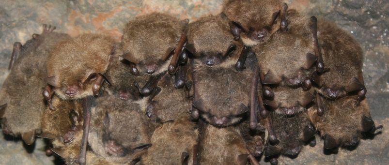
Bats infected by white nose syndrome.
Thursday, March 28, 2013
Tuesday, March 19, 2013
WNS and Culture samples
Like histology, culture samples require skin tissue for testing. Culture samples require a 1.5cmx1.5cm piece of wing tissue from the bat. This sample is then placed on a dextrose medium containing chloramphenicol and gentamycin. Plates are incubated at 45°F for 10-30 days and examined every 1-3 days for traces of G. destructans. Biologists look for curved single-celled conidia when looking at samples under a microscope. Fungal cultures are more complicated than histology tests because of the nonsterile nature of bat wings. Tissues for culture tests may be removed from the muzzle, wing, or ear tissues, though wing tissue is preferred. Like histology tests, results take a long time to yield conclusive data and sample sizes are so big that the bat being studied can not survive once a culture sample has been removed. The following image is what biologists look for when determining if bats have been infected with WNS.


Saturday, March 16, 2013
Histology and WNS
Histology is considered to be the standard for determining White Nose Syndrome in bat species in the United States and Europe. To determine if a bat has WNS, a 1.5cmx3.0cm piece of wing membrane must be removed from a bat then cut and prepared. Cross sections of the rolled wing skin are then examined microscopically for the presence of the fungus associated with White Nose Syndrome, Geomyces destructans. Biologists specifically look for conidia in conjunction with fungal hyphae, cup-like epidermal erosions, ulsers and damage to underlying connective tissues. This method is often considered to be labor intensive and time consuming, with results taking more than a week to acquire. A downside to histology methods is that this technique, while sensitive and reliable, requires specialized training for the interpretation of results. The largest downfall with this method is that it is invasive to the animal being tested and requires a large amount of tissue to be removed from the bat, often a piece too large to allow the bat to remain living. Below are a few pictures of histology test results determining if a bat has WNS.

 (A) Direct lactophenol cotton blue mount prepared from skin scrape taken
from the muzzle of a little brown bat from Graphite Mine on April 6,
2008 revealed fungal hyphae and curved conidia, bar 10 µm. (B) Control,
[Bi] and infected muzzle tissue section [Bii] stained with PAS revealed
epidermal colonization by fungal hyphae and spores; the sample was from a
little brown bat from Williams Hotel Mine on March 27, 2008. Notably, a
few neutrophils are present in the underlying dermis (arrows), bar 10
µm. Bacteria are also seen in this sample (C). SEM photomicrograph of
muzzle sample from bat from Williams Hotel Mine showing characteristic
curved conidia and septate hyphae spread over bat skin tissues. Note
heavy fungal growth with profuse curved conidia covering the skin and
hair shaft (Ci, muzzle, bar 100 µm; Cii, higher magnification of a
portion of muzzle, bar 10 µm; Ciii & Cvi, higher magnifications, bar
10 µm).
(A) Direct lactophenol cotton blue mount prepared from skin scrape taken
from the muzzle of a little brown bat from Graphite Mine on April 6,
2008 revealed fungal hyphae and curved conidia, bar 10 µm. (B) Control,
[Bi] and infected muzzle tissue section [Bii] stained with PAS revealed
epidermal colonization by fungal hyphae and spores; the sample was from a
little brown bat from Williams Hotel Mine on March 27, 2008. Notably, a
few neutrophils are present in the underlying dermis (arrows), bar 10
µm. Bacteria are also seen in this sample (C). SEM photomicrograph of
muzzle sample from bat from Williams Hotel Mine showing characteristic
curved conidia and septate hyphae spread over bat skin tissues. Note
heavy fungal growth with profuse curved conidia covering the skin and
hair shaft (Ci, muzzle, bar 100 µm; Cii, higher magnification of a
portion of muzzle, bar 10 µm; Ciii & Cvi, higher magnifications, bar
10 µm).
The second picture and description were taken from an article that can be found at http://www.plosone.org/article/info%3Adoi%2F10.1371%2Fjournal.pone.0010783.


The second picture and description were taken from an article that can be found at http://www.plosone.org/article/info%3Adoi%2F10.1371%2Fjournal.pone.0010783.
Wednesday, March 13, 2013
Detecting bats with WNS
It almost seems that White Nose Syndrome is being discovered in another state or province daily. Recent news has stated that WNS has been found in Georgia and North Carolina now. There is a lot of talk about where it is discovered, but not much on HOW. How do biologists determine if a bat has contracted this disease that is killing almost 100% of populations that it infects? The answer isn't so simple. Biologists first identified WNS from a picture from Howes cave near Albany New York. Today, a photograph isn't enough evidence to suggest an individual is infected. Biologists typically use one of 3 methods to determine if a bat has WNS. These methods include histology, culture samples, and the use of polymerase chain reaction (PCR). Tape has been used in the past to remove fungal particles from the wings and faces of those presumed infected, however, the three aforementioned methods are considered to be the definite methods for determining infection. Depending on the method used, results can take between hours and weeks before biologists have conclusive answers. Both histology and culture methods require weeks for definitive results but PCR methods allow for rapid answers, sometimes with hours. Over the course of the next week I intend to go into further detail regarding each of these methods for tracking and determining if populations have WNS. I also intend to discuss how soil samples from caves have lead to knowledge about WNS.


Monday, March 4, 2013
USGS Update
Several weeks ago I uploaded a map provided by the USGS regarding the prevalence of WNS across the country.. Today they issued a new map....
 Note the section in Illinois and Canada that now have confirmed cases of WNS that were previously not on the map at all. It also appears that confirmed cases have now been found in Kentucky and Tennessee as well.
As White Nose Syndrome spreads across the country effecting migratory species in their hibernacula, you will see a decrease in the number of bats around your homes during the summer months once they have returned to their summer roosts. You know what that means? More mosquitoes and pests that are eaten by bats as they feed at night. Did you know that WNS only infects insectivorous bat species int he U.S.? This will inevitably have an impact on the number of pests around our homes and will result in the increased use of pesticides that pose other damaging issues to our environment.

As White Nose Syndrome spreads across the country effecting migratory species in their hibernacula, you will see a decrease in the number of bats around your homes during the summer months once they have returned to their summer roosts. You know what that means? More mosquitoes and pests that are eaten by bats as they feed at night. Did you know that WNS only infects insectivorous bat species int he U.S.? This will inevitably have an impact on the number of pests around our homes and will result in the increased use of pesticides that pose other damaging issues to our environment.
Subscribe to:
Posts (Atom)